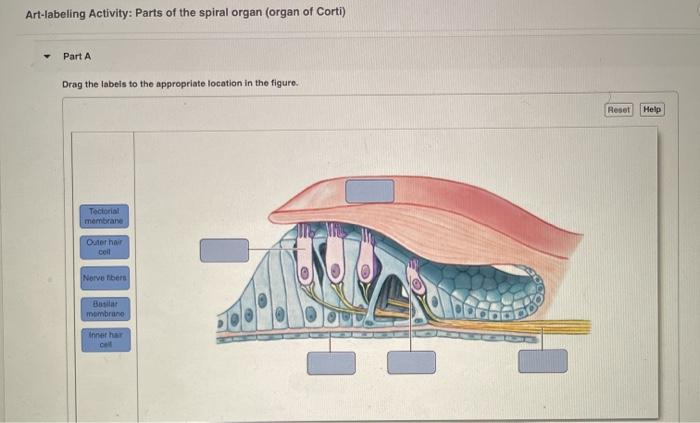
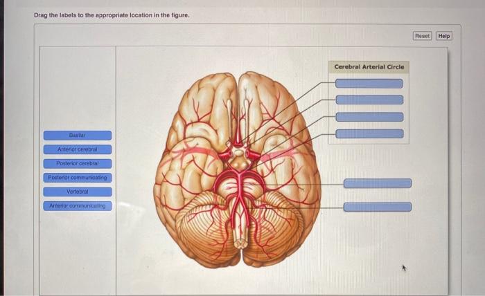
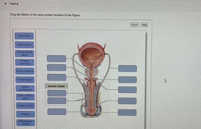
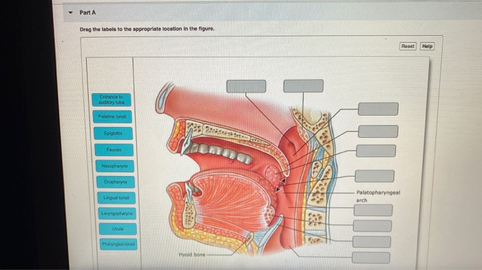
41 drag the labels to the appropriate location in the figure.
Solved Art-labeling Activity: Neuron Structure 6 of 36 - Chegg Art-labeling Activity: Neuron Structure 6 of 36 Review Part A Drag the labels to the appropriate location in the figure. Axon Nucleus Synaptic terminals Microfibrils and microtubules CO on Coll body II. Mitochondrion Dendrites Nucleolus Submit Request Answer Solv cheo This problem has been solved! AP 1 (Histology) Flashcards | Quizlet Drag the appropriate labels to their respective targets. Use labels of Group 1 for the tissues and labels of Group 2 for the structures. Labels can be used once or more than once. Identify the cells found within this tissue. Fibroblasts Identify the type and tissue shown in the image Spongy Bone Connective Tissue
Drag the labels to the appropriate location in the figure. Drag the labels to their appropriate locations in the figure. First, drag labels to targets (a) and (b) to indicate whether these environments are hydrophilic or hydrophobic. Next, drag the phospholipid layers to targets (c) and (d) to indicate how they are oriented in the plasma membrane.
Drag the labels to the appropriate location in the figure.
Mastering Biology: Chapter 7 Flashcards | Quizlet Drag the labels to their appropriate locations in the figure. First, drag labels to targets (a) and (b) to indicate whether these environments are hydrophilic or hydrophobic. Next, drag the phospholipid layers to targets (c) and (d) to indicate how they are oriented in the plasma membrane. 1.4 Anatomical Terminology - Anatomy & Physiology Figure 1.4.1 - Regions of the Human Body: The human body is shown in anatomical position in an (a) anterior view and a (b) posterior view. The regions of the body are labeled in boldface. A body that is lying down is described as either prone or supine. Part A Drag the labels to the appropriate location in the figure ANSWER ... Part A Drag the labels to the appropriate location in the figure ANSWER Correct. Part a drag the labels to the appropriate location in. School Miami Dade College, Miami; Course Title ANAT 260; Uploaded By cami032312. Pages 10
Drag the labels to the appropriate location in the figure.. drag the labels to the appropriate location in the figure. [Solved ... drag the labels to the appropriate location in the figure. Answer to question 1 Answer to question 2 Step 1 Epithelial tissue- is one of the four basic types of tissue found in an animal. Epithelial tissue lines the outer surfaces of organs and blood vessels throughout the body and also the inner surfaces of cavities of internal organs. Step 2 Answered: Drag the labels to the appropriate… | bartleby Transcribed Image Text: Part A Drag the labels to the appropriate location in the figure. Reset Help Simple squamous epithelium Simple columnar epithelium Simple cuboidal epithelium Pseudostratified ciliated columnar epithelium Transitional epithelium Stratified squamous epithelium O Typę here to search. Part A Drag the labels to the appropriate location in the figure ANSWER ... Part A Drag the labels to the appropriate location in the figure ANSWER Correct. Part a drag the labels to the appropriate location in. School Miami Dade College, Miami; Course Title ANAT 260; Uploaded By cami032312. Pages 10 1.4 Anatomical Terminology - Anatomy & Physiology Figure 1.4.1 - Regions of the Human Body: The human body is shown in anatomical position in an (a) anterior view and a (b) posterior view. The regions of the body are labeled in boldface. A body that is lying down is described as either prone or supine.
Mastering Biology: Chapter 7 Flashcards | Quizlet Drag the labels to their appropriate locations in the figure. First, drag labels to targets (a) and (b) to indicate whether these environments are hydrophilic or hydrophobic. Next, drag the phospholipid layers to targets (c) and (d) to indicate how they are oriented in the plasma membrane.
Solved Drag the labels to the appropriate location in the ...

Solved Part A Drag the labels to the appropriate location in ...
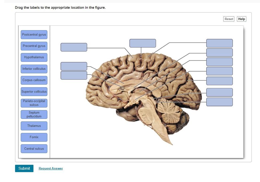
Solved Drag the labels to the appropriate location in the ...
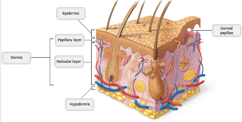
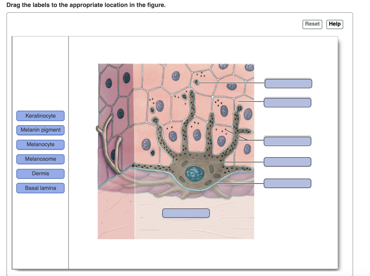
A&P Chapter 5 The Integumentary System Flashcards - Easy ...
Solved Part A Drag the labels to the appropriate location in ...

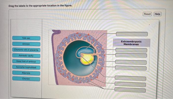
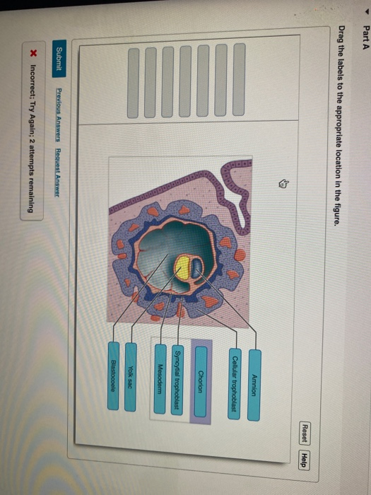
SOLVED: igure Walkthrough: The Human Lite Cycle Drag the ...

Chapter 1 Flashcards | Quizlet
Solved Drag the labels to the appropriate location in the ...
![Expert Answer] Drag labels to the appropriate locations in ...](https://us-static.z-dn.net/files/dd9/363dc6f0791b99410cbee879fdbd9356.png)
Expert Answer] Drag labels to the appropriate locations in ...

Lc 14 HW.pdf - Lc 14 HW Due: 7:59am on Tuesday, May 19, 2020 ...

Mastering Quiz 1: Chapter 16 Flashcards | Quizlet

Lab 9 HW Flashcards | Quizlet

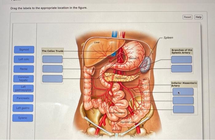
digestive systems (my lab and mastering) Flashcards | Quizlet
Solved Drag the labels to the appropriate location in the ...
Solved Drag the labels to the appropriate location in the ...

Ch 5-7 Lab A&P Mastering Flashcards | Quizlet

Ch 5-7 Lab A&P Mastering Flashcards | Quizlet
Answered: Drag the labels to the appropriate… | bartleby
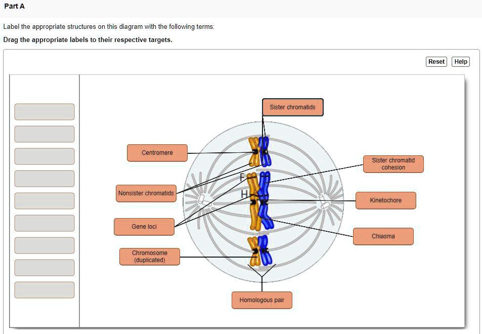
SOLVED: Part A Label the appropriate structures on this ...

Lab 12 mastering a&p Flashcards | Quizlet

Chapter 1 Flashcards | Quizlet
Solved Part A Drag the labels to the appropriate location in ...

Drag each label to the correct location on the diagram. Each ...
Solved] Part A The figure shows the global water cycle. Drag ...
Solved] Using the drag-and-drop environment, drag the labels ...
Solved Drag the labels to the appropriate location in the ...
Solved Drag the labels to the appropriate location in the ...
Solved Drag the labels to the appropriate location in the ...
Solved Drag the labels to the appropriate location in the ...
![ANSWERED] 3 Drag each label to the correct location on the ...](https://media.kunduz.com/media/sug-question-candidate/20230102032108057038-4073814.jpg)
ANSWERED] 3 Drag each label to the correct location on the ...
Solved Part A Drag the labels to the appropriate location in ...
Solved] IF you can help me answer this I would appreciate it ...

Lab 2--exercise 3&4 Flashcards | Quizlet
Solved Part A Drag the labels to the appropriate location in ...

On this map of the world, drag the label for each type of ...

Answers to mastering biology drag the labels to their ...
Solved Part A Drag the labels to the appropriate location in ...
Solved Part A Drag the labels to the appropriate location in ...
Solved] e questions. Label each component of a nucleotide ...

Chapter 14 Flashcards | Quizlet
![Answered] Drag the labels to the appropriate locations on ...](https://us-static.z-dn.net/files/d74/87a7a59e3181c46ccbe8bef4641d3256.png)
Answered] Drag the labels to the appropriate locations on ...
Post a Comment for "41 drag the labels to the appropriate location in the figure."