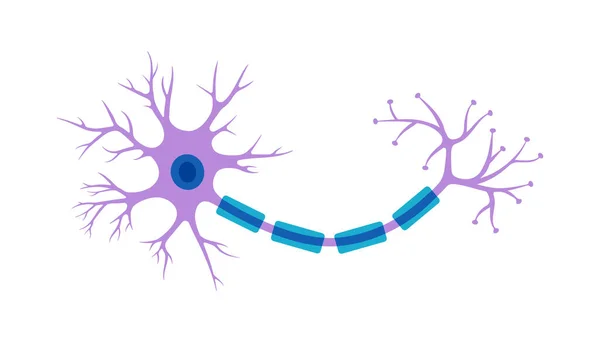
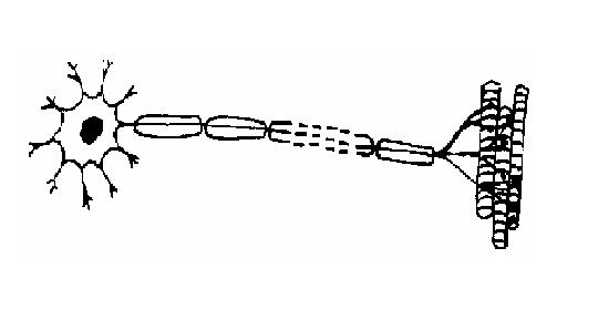
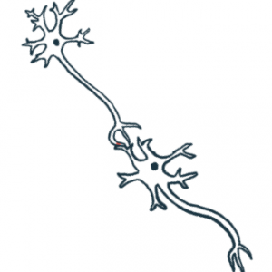
42 neuron diagram unlabeled
Human Ear Diagram - Bodytomy The Structure of Human Ear. Helix: It is the prominent outer rim of the external ear. Antihelix: It is the cartilage curve that is situated parallel to the helix. Crus of the Helix: It is the landmark of the outer ear, situated right above the pointy protrusion known as the tragus. Auditory Ossicles: The three small bones in the middle ear ... Neuroscience for Kids - Fill In #1 Use the words from the list below to label the following diagram of a neuron in the lines provided. ... Below is a list of different parts of a neuron.
Neuron Nerve Cell Diagram Blank Sketch Coloring Page The nervous system sends messages from nerve endings to the brain and from the brain to cells, tissues, and organs. Cells of the nervous system sometimes secret. Bubakids askabiologist asu Edu Activities Coloring Animal Cell for preschool, kindergarten and elementary school children to print and color.

Neuron diagram unlabeled
neuron diagram worksheet neuron diagram blank worksheet brain cell unlabeled nervous system anatomy nerve labeling neurons printable label body human motor central parts Neuron Label neuron label nervous anatomy system nerve worksheet cell diagram key answer neurons biology human concept map brain coloring labeling physiology Labeled Neuron Diagram | Science Trends Neurons form the bulk of all nervous tissue and are what allow nervous tissue to conduct electrical signals that allow parts of the body to communicate with each other. Neurons are the cells that are responsible for receiving sensory input from the outside world, sending motor commands to move parts of the body, forming memories in the brain, and more. neuron cell labeled neuron diagram cell complete nervous system components neurons brain biology human physiology anatomy nerve membrane parts cells potential science neurone. ... neuron diagram blank worksheet brain cell unlabeled nervous system anatomy nerve neurons label printable human labeling body motor central parts. Chapter 8, Page 8 - HistologyOLM 4.0 ...
Neuron diagram unlabeled. Simple nephron diagram - Healthiack Best viewed on 1280 x 768 px resolution in any modern browser. Simple nephron diagram 1239. Simple nephron diagram 1243. Simple nephron diagram 1244. Simple nephron diagram 1245. Simple nephron diagram 1248. Simple nephron diagram 1253. Simple nephron diagram 1256. Simple nephron diagram 1264. neuron labeled diagram 34 Draw And Label The Parts Of A Neuron - Label Design Ideas 2020 dandelionsandthings.blogspot.com. neuron parts label draw neurons lab. Behind The Science: The Anatomy Of A Neuron blog.eyewire.org. neuron anatomy cell behind science structure biology molecular lodish berk freeman sl 4th section 2000 et al edition york neuron diagram labeled brain diagram human unlabeled worksheet eye system nervous labeling anatomy worksheets label worksheeto via muscular lab Neural Signal Transmission signal transmission neural neuron Anatomy/Nervous System - Wiki - Scioly.org scioly.org nervous neuron scioly Answer: A = Neuron Cell Body, B = Glial Cell Body, C = Axons Motor neuron unlabeled. - Alila Medical Media Image size: 31.2 Mpixels (89.4 MB uncompressed) - 5000x6250 pixels (16.6x20.8 in / 42.3x52.9 cm at 300 ppi) Published in: Brain and Nervous System Images , Neurology Images & Videos, Cell, Molecular Biology & Genetics Images. Powered by PhotoDeck.
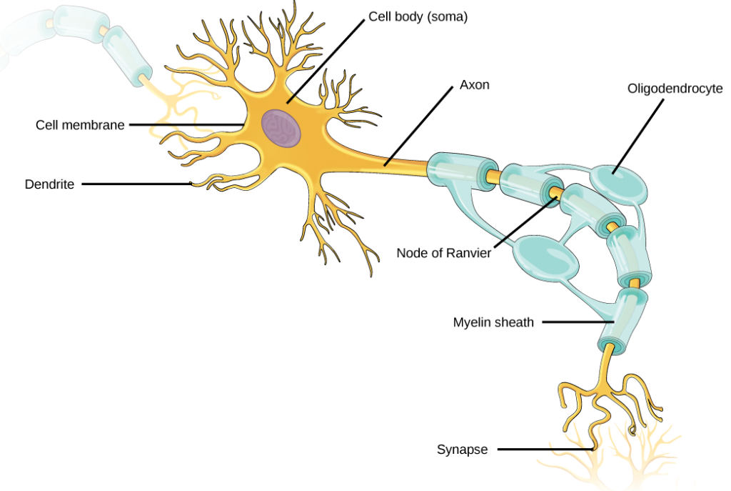
General Structure of a Neuron (Nerve Cell) | GetBodySmart General Structure of a Neuron (Nerve Cell) Start Quiz. Learn this topic from scratch or practice what you already know with these interactive spaced repetition-inspired anatomy quizzes. Learn anatomy faster and. remember everything you learn. Start Now. <. Nervous System - Label the Neuron Nervous System - Label the Neuron Nervous System - Neuron: Nerve Cell Name: Choose the correct names for the parts of the neuron. (1) (2) (3) (4) (5) (6) This neuron part receives messages from other neurons. (7) This neuron part sends on messages to other neurons. (8) This neuron part gives messages to muscle tissue. Question Video: Recalling the Ion Which Generates an Action ... - Nagwa The diagram provided shows a simplified unlabeled outline of a cholinergic synapse. The influx of which ion generates an action potential in the postsynaptic neuron? (A) Calcium, (B) oxygen, (C) sodium, (D) potassium, (E) hydrogen. The Respiratory System Diagram - Liveworksheets The Respiratory System Diagram This worksheet will help students learn the repsiratory system ID: 2737173 Language: English School subject: health Grade/level: 12 Age: 16-18 Main content: Diagram of the repsiratory system Other contents: label the respiratory system Add to my workbooks (0)
neuron diagram blank BasicStructure Of Neurons: neuron diagram label anatomy cell brain nerve blank unlabeled neurons structure system nervous body worksheet enchanted learning coloring cells axon Motor Neuron, Detail And Accurate,non-labeled Vs Stock Vector neuron motor labeled accurate non vs royalty illustration A Labelled Diagram Of Neuron with Detailed Explanations - BYJUS Diagram Of Neuron Diagram Of Neuron A neuron is a specialized cell, primarily involved in transmitting information through electrical and chemical signals. They are found in the brain, spinal cord and the peripheral nerves. A neuron is also known as the nerve cell. Neuron Diagram & Types | Ask A Biologist - Arizona State University Multipolar neurons have one axon and many dendritic branches. These carry signals from the central nervous system to other parts of your body such as your muscles and glands. Unipolar neurons are also known as sensory neurons. They have one axon and one dendrite branching off in opposite directions from the cell body. Tactile Neuron Diagram (Unlabeled) by trynne - Thingiverse Download files and build them with your 3D printer, laser cutter, or CNC. Thingiverse is a universe of things.
The Nervous System | Biology - Quizizz
Simple Diagram Of Nephron Each nephron is made up of two parts: a renal corpuscle and renal tubules Schematic diagram of the nephron demonstrating the site of action of diuretics. The nephron is the microscopic structural and functional unit of the kidney. It is composed of a Diagram (left) of a long juxtamedullary nephron and (right) of a short cortical nephron. .
Ranvierov čvor - Wikipedia
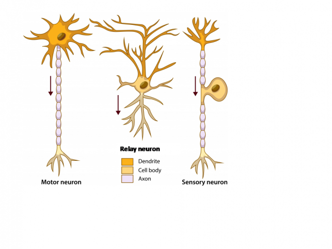
Sensory Neuron - The Definitive Guide | Biology Dictionary Neurons are cells of the nervous system that can transmit electrical impulses to facilitate communications between the brain and the rest of the body. There are three main types of neurons: sensory neurons, relay neurons, and motor neurons.

Neuron, Motor Neuron, Sistem Saraf gambar png
Neurons (With Diagram) - Biology Discussion A neuron consists of main cell body and cytoplasmic processes arising from it. ADVERTISEMENTS: (i) Cell body (= Cyton or Soma): It varies in size and form. It may be up to 13.5 µm in diameter and may be irregular, spherical, oval, rounded, star-shaped or pyramidal. Like a typical cell it consists of cytoplasm, nucleus and cell membrane.

Single-Neuron-Based AI Reacts to the Human Voice | Electronic ...
Nervous System Worksheet - WikiEducator 8. The diagram below shows a section of a dog's brain. Add the labels in the list below and, if you like, colour in the diagram as suggested. Cerebellum - blue; Spinal cord - green; Medulla oblongata - orange; Hypothalamus - purple; Pituitary gland - red; Cerebral hemispheres - yellow. 9. Match the descriptions below with the terms in the list.

Neuron Model
Sensory Neuron Diagram Illustrations & Vectors - Dreamstime Labeled diagram of the Neuron, nerve cell that is the main part of the nervous system. Abstract grey mesh background Human nervous system medical vector illustration diagram with parasympathetic and sympathetic nerves and connected inner organs.

Neuron Structure Diagram Silhouette @ Silhouette.pics
Neuron Nerve Cell Diagram Blank Sketch Coloring Page Jun 8, 2016 - This Pin was discovered by Shelly Peacock. Discover (and save!) your own Pins on Pinterest.

Label Neuron Anatomy Printout - EnchantedLearning.com
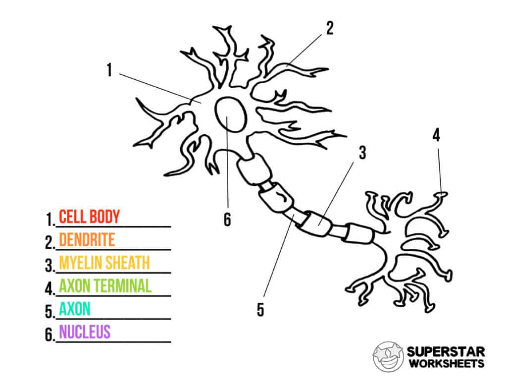
Oversized Neuron & Types of Neurons Diagrams - Incl Notes & Bonus ... The structures of the neuron is a challenging thing to learn when studying the Nervous System. These oversized diagrams focus on the most important structures of the neuron that every student needs to know. Here is the list of the structures this resource covers: List of Structures on Neuron Diagram (8) Axon. Axonal Terminals. Cell Body. Dendrite

Nerve Impulse | CK-12 Foundation
Neuron Anatomy - Worksheet Activity - Ask A Biologist Neuron Anatomy Activity. The parts of the neuron have been labeled. Your challenge is to write the correct name for each part and explain what it does.

Grab your free printable neuron cell worksheets from ...
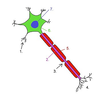
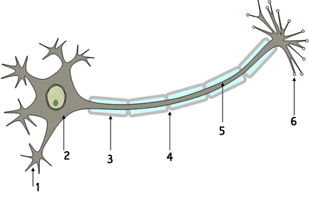
Neuron Diagram Unlabeled neuron, (1). axon, cell body, dendrites, nucleus, terminal. Unlabeled diagram of a motor neuron (try labeling: axon, dendrite, cell body, myelin, nodes of Ranvier, motor end plate).Read the definitions, then label the neuron diagram below. axon - the long extension of a neuron that carries nerve impulses away from the body of the cell.

assignt.htm
Neuron Diagram - Printable Worksheet - BrainFrame-Kids Free printable neuron diagram worksheet.

I seriously think ms. Farsii made us label this exact nerve ...
car diagram unlabeled Free cars movie cliparts, download free cars movie cliparts png images. Nervous system function parts structures nerve brain diagram neuron messages anatomy. Arteries major systemic unlabeled carotid artery unlabelled blood vessels neck clipart transparent body label ekg common internal anatomy webstockreview skeleton car diagram unlabeled
The Neuron
neuron with labeled parts brain diagram human unlabeled worksheet eye system nervous labeling anatomy worksheets label worksheeto via muscular lab. Neuron Anatomy - Anatomy Drawing Diagram sen842cova.blogspot.com. neuron neurons. Science - 8th Grade: Neuron sciencejl.blogspot.com. 8th grade neuron science figure nervous system. Auditory On A Diagram Of A Neuron ...

Neuron Labeled Diagram - ClipArt Best
labeled diagram of neuron labeled diagram of neuron Structure of a typical motor neuron we have 9 Pictures about Structure of a typical motor neuron like Nerve Cell Diagram Labeled 2019 - 101 Diagrams, The Anatomy and Physiology of Animals/Nervous System Worksheet and also Neuron With Axon Clip Art at Clker.com - vector clip art online. Read more:

Chapter 26 The Nervous System - 26 Test Prep for AP Courses
anatomy of a neuron worksheet Neuron diagram blank worksheet brain cell unlabeled nervous system anatomy nerve labeling neurons printable label body human motor central parts. Neuron worksheet diagram nervous system blank cell nerve label elementary synapse science worksheeto body brain folder via games file biology ... brain diagram human unlabeled worksheet eye system ...

neuron | North Central AP Psychology
Neuron Diagram Teaching Resources | Teachers Pay Teachers Neurons & Neurotransmitters Reading, Neuron Diagram, Transmitter Chart & Skit. by. Learning the Social Sciences. 4.9. (7) $1.75. Zip. This item contains a one-page reading explaining neurotransmitters and neurons. It discusses neurotransmission, the axon terminal button, vesicles, the axon, resting potential, action potential, and more.

SVG > cell neurology intelligence neuron - Free SVG Image ...
blank brain diagram inside Ap Psych Brain Diagram Unlabeled - Motor Neuron Not Labeled - Free. 9 Pics about Ap Psych Brain Diagram Unlabeled - Motor Neuron Not Labeled - Free : Brain Blank Diagram - ClipArt Best, Brain Sagittal Section H | Free Images at Clker.com - vector clip art and also Brain Sagittal Section H | Free Images at Clker.com - vector clip art.
Neuron Diagrams
neuron cell labeled neuron diagram cell complete nervous system components neurons brain biology human physiology anatomy nerve membrane parts cells potential science neurone. ... neuron diagram blank worksheet brain cell unlabeled nervous system anatomy nerve neurons label printable human labeling body motor central parts. Chapter 8, Page 8 - HistologyOLM 4.0 ...
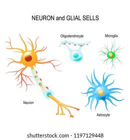
120 Neuroglia Images, Stock Photos & Vectors | Shutterstock
Labeled Neuron Diagram | Science Trends Neurons form the bulk of all nervous tissue and are what allow nervous tissue to conduct electrical signals that allow parts of the body to communicate with each other. Neurons are the cells that are responsible for receiving sensory input from the outside world, sending motor commands to move parts of the body, forming memories in the brain, and more.
Describe the Nervous System Worksheet - EdPlace
neuron diagram worksheet neuron diagram blank worksheet brain cell unlabeled nervous system anatomy nerve labeling neurons printable label body human motor central parts Neuron Label neuron label nervous anatomy system nerve worksheet cell diagram key answer neurons biology human concept map brain coloring labeling physiology
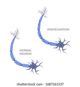
Healthy Damaged Neuron Diagram Demyelination Neuron Stock ...

Neuron Nerve Cell Diagram Blank Sketch Coloring Page ...
Neuron Cell Worksheets - Superstar Worksheets
Nervous System Review 9-1 to 9.10
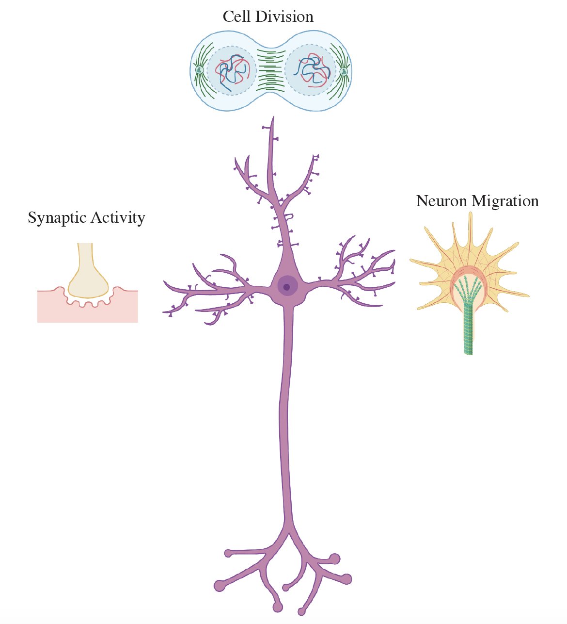
Dr Mohammad Mofatteh on Twitter: "Regulation of gene ...
Histology of neurons: Morphology and types of neurons | Kenhub
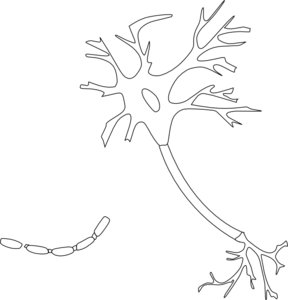
neuron diagram unlabeled - Clip Art Library
Axon Vektor Stok, Ilustrasi Axon Bebas Royalti - Halaman 8 ...
7.2 – Resting, Graded and Action Potential – Introductory ...
Solved] Please see attachments for details | Course Hero

Neuron Anatomy Hand Drawn Vector Illustration Like Woodblock ...

The Neuron Diagram Diagram | Quizlet
Nervous System Worksheet - WikiEducator

Lesson Worksheet:Neurons | Nagwa

Sensory Neuron - The Definitive Guide | Biology Dictionary

Structure of A Typical Neuron stock vectors and illustrations

Sensory Neuron
MS Gray Matter Loss in Spine Crucial, But Difficult, Marker ...

n Illustration of neurotransmitters being released from the ...

Label The Neuron Clip Art at Clker.com - vector clip art ...

Q3 Given below is the diagram of a neuron Name the parts ...
Frontiers | Carbonic Anhydrases as Potential Targets Against ...

Which one of the following pairs is incorrect?
Post a Comment for "42 neuron diagram unlabeled"