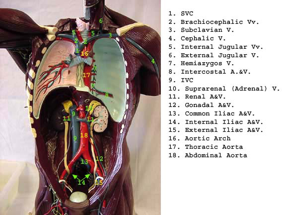
39 label thoracic cavity
1.4E: Body Cavities - Medicine LibreTexts The thoracic cavity is lined by two types of mesothelium, a type of membrane tissue that lines the ventral cavity: the pleura lining of the lungs, and the pericadium lining of the heart. Abdominopelvic. The abdominoplevic cavity is the posterior ventral body cavity found beneath the thoracic cavity and diaphragm. It is generally divided into ... [Solved] Label the structures of the thoracic cavity | Course Hero The thoracic cavity is a large, hollow space in the chest that contains the lungs, heart, and other organs. The cavity is divided into two parts: the pleural cavity and the pericardial cavity. Pleural cavity is lined with a thin layer of tissue called the pleura. The pericardial cavity is the space between the two layers of the pericardium
Body Cavities and Organs with Labeled Diagram - AnatomyLearner The thoracic cavity of an animal is cone-shaped and laterally compressed. In addition, the abdominal cavity of the animal is the largest cavity that extends from the diaphragm to the pelvic inlet. The vertebral cavity contains the spinal cord and the roots of the spinal nerves of an animal.

Label thoracic cavity
Thoracic Cage Labeling Quiz - PurposeGames.com This is an online quiz called Thoracic Cage Labeling There is a printable worksheet available for download here so you can take the quiz with pen and paper. Your Skills & Rank Total Points 0 Get started! Today's Rank -- 0 Today 's Points One of us! Game Points 13 You need to get 100% to score the 13 points available Actions Oxytetracycline (Terramycin®, Liquamycin®) for Dogs and Cats Jul 16, 2015 · Oxytetracycline is effective against a wide range of bacteria as well as one-celled (protozoa) organisms. It is effective against bacteria that infect the eyes, oral cavity, bone, respiratory tract, sinuses and blood cells. Oxytetracycline is a prescription drug and can only be obtained from a veterinarian or by prescription from a veterinarian. Anatomy Chapter 1: Labeling Thoracic Cavity Diagram | Quizlet The cavities surrounding each lung parietal pleura The aspect of the pleura that does not touch the surface of the lung visceral pleura The aspect of the pleura that covers the external surface of the lung The thoracic cavity can be subdivided into... 1. mediastinum 2. left and right pleural cavities 3. pericardial cavity
Label thoracic cavity. Costodiaphragmatic recess - Wikipedia The costodiaphragmatic recess, also called the costophrenic recess or phrenicocostal sinus, is the posterolateral fringe of the pleural space, a potential space around the lung inside the pleural cavity.It is located at the acutely angled junction ("reflection") between the costal and diaphragmatic pleurae, and is interpreted two-dimensionally on plain X-rays as the … Thoracic and mediastinal lymph nodes and lymphatics | Kenhub Overview The thorax is the region of the body extending from the base of the neck and thoracic inlet (the latter being at the supraclavicular fossae) to the diaphragm (marked anteriorly by the xiphisternal joint).. Within the thoracic cavity is the mediastinum.The mediastinum is the region of the thorax between the lungs.It extends from the level of the first rib, superiorly, to the diaphragm ... Labeled Diagram of the Human Lungs - Bodytomy Human lungs are located in the thoracic cavity or chest and are enclosed within the rib cage. The two lungs are situated on either sides of the heart and are pinkish in color, especially at a young age. Exposure to the atmosphere and polluted air eventually gives rise to mottled patches, which tint the lungs gray in color. Major Body Cavities, Their Subdivisions And Membranes. The thoracic cavity is guarded by the rib cage and contains the heart and lungs. The abdominopelvic cavity is subdivided into a superior abdominal cavity and an inferior pelvic cavity, however, there is no structural separation between them. To visualize the separation, think of a transverse plane passing through the body just superior to the ...
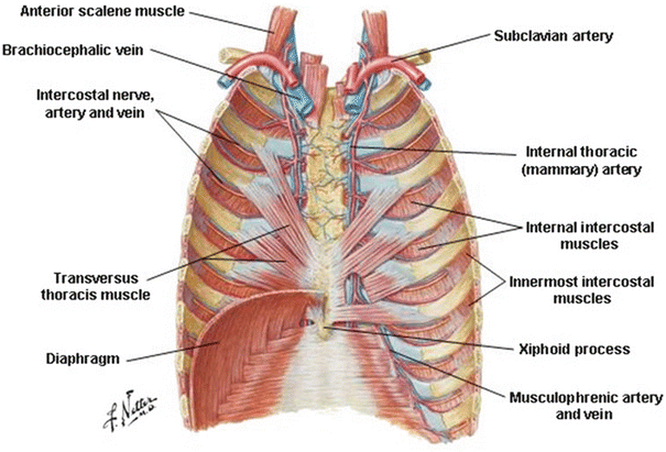
Answered: Correctly label the muscles of the… | bartleby The thoracic artery is also known as the internal mammary artery. It supplies the breasts and the anterior chest wall. There are two internal arteries, the right and left artery, which are situated anterior to the chest wall… Article Respiratory System arrow_forward Label the Body Cavities Diagram | Quizlet Start studying Label the Body Cavities. Learn vocabulary, terms, and more with flashcards, games, and other study tools. Fetal Pig Dissection - Virtual Anatomy & Diagrams | HST Abdominal Cavity. 1. The largest organ in the abdominal cavity is by far the liver, just below the diaphragm (the flap of muscle separating the abdominal from the thoracic cavity). Notice the umbilical vein connecting the umbilical cord with the liver. Cut this vein so you can lay the umbilical cord back between the pig's hind legs. 2010 ACCF/AHA/AATS/ACR/ASA/SCA/SCAI/SIR/STS/SVM 30-07-2022 · Familial aggregation studies of patients referred for repair of thoracic aortic aneurysm and dissection that did not have a genetic defect have indicated that between 11% and 19% of these patients have a first-degree relative with thoracic aortic aneurysms and dissection. 127,130 Patients with a family history of thoracic aortic aneurysm and dissection present at a …
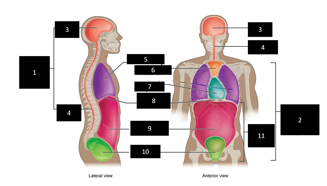
Solved Correctly label the following area of the thoracic - Chegg Expert Answer 100% (18 ratings) Transcribed image text: Correctly label the following areas of the thoracic cavity in the newborn and the adult. Pituitary land Diaphragm Liver Lung Trachea Adrenal gland Thyroid Heart Pineal gland Thymus (a) Newborn (b) Adult Previous question Next question Anatomy, Thorax - StatPearls - NCBI Bookshelf - National Center for ... Last Update: July 31, 2021. Pleural cavities - lungs and pleura Superior mediastinum - great vessels, trachea, esophagus, vagus nerve, phrenic nerve, sympathetic nerves, thoracic lymphatic duct, thymus Anterior mediastinum - connective tissue, thymus, and lymph nodes Middle mediastinum - heart, roots of great vessels, phrenic nerve, and pericardium Solved Award: 0.76 points Label the structures of the - Chegg Science. Anatomy and Physiology. Anatomy and Physiology questions and answers. Award: 0.76 points Label the structures of the thoracic cavity. Parietal pleura Visceral pleura Pleural cavity Parietal pericardium Visceral pericardium Pericardial cavity Reset Zoom. Question: Award: 0.76 points Label the structures of the thoracic cavity. 685 Thoracic cavity Images, Stock Photos & Vectors - Shutterstock 685 thoracic cavity stock photos, vectors, and illustrations are available royalty-free. See thoracic cavity stock video clips Image type Orientation People Artists Sort by Popular Healthcare and Medical Biology Anatomy Recreation/Fitness lung thorax thoracic cavity chest radiograph radiography human body Next of 7
Ventral Body Cavity | Subdivisions, Organs, & Diagram Video
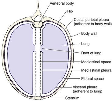
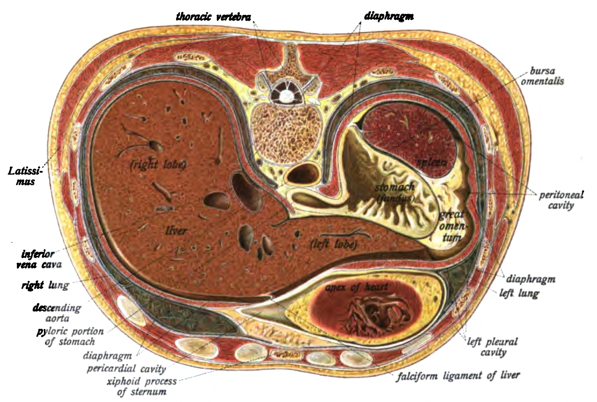
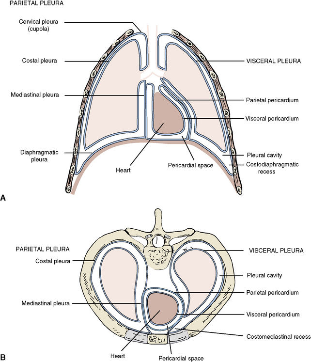
Thoracic Cavity - Definition & Organs of Chest Cavity - Biology Dictionary The thoracic cavity is actually composed of three spaces each lined with mesothelium, a special film-like tissue that separates vital organs. The pleural cavities surround the lungs, while the pericardial cavity surrounds and protects the heart. These tissues in the thoracic cavity can be seen in the image below.

Location of the heart within the mediastinum of the thoracic ...
Color Diagrams of Insect Organs and Internal Structures 17-01-2019 · Insects don't have veins or arteries, but they do have circulatory systems. When blood is moved without the aid of vessels, the organism has an open circulatory system. Insect blood, properly called hemolymph, flows freely through the body cavity and makes direct contact with organs and tissues.
1,050 Costal cartilage Images, Stock Photos & Vectors ...
Thorax: Anatomy, wall, cavity, organs & neurovasculature | Kenhub Thoracic wall The first step in understanding thorax anatomy is to find out its boundaries. The thoracic, or chest wall, consists of a skeletal framework, fascia, muscles, and neurovasculature - all connected together to form a strong and protective yet flexible cage.. The thorax has two major openings: the superior thoracic aperture found superiorly and the inferior thoracic aperture ...
Thorax: Anatomy, wall, cavity, organs & neurovasculature | Kenhub
Membranes and cavities - Human Anatomy - GUWS Medical Figure 2.1 Label the major body cavities. (cavity) (canal or cavity) Dorsal cavity. Figure 2.2 Label the smaller cavities and sinuses within the head. Figure 2.2 Label the smaller cavities and sinuses within the head. Figure 2.3 Label the thoracic membranes and cavities in (a) and the abdominopelvic membranes and cavity in (b) as shown in these ...
Thoracic Cavity | Veterian Key
Thoracic cavity | Whitman College Thoracic cavity Section Navigation In this photo, we can see the heart contained within in its transparent pericardial membrane. Also found inside the thoracic cavity are the right and left lungs, which are on either side of the heart. Also note the thymus gland, which in many young mammals can be found in the throat and the thoracic cavity.

Label the organs 1. brain 2. Thyroid gland 3. Trachea 5 ...
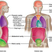
Anatomical Body Planes | Science Trends Examples of these anatomical terms used to label body structures include the axial skeleton, the median cerebral artery, ... the vertebral cavity, the thoracic cavity, and the abdominopelvic cavity. The dorsal cavity is one long continuous cavity that houses portions of the central nervous system including the spinal cord and brain.
Body Cavities Labeling
right posterior oblique position definition how to draw contour lines on a grid; always check up on your friends Menu Toggle. weather auburn ca hourly; picasso immersive exhibition vancouver; copenhagen natural disasters
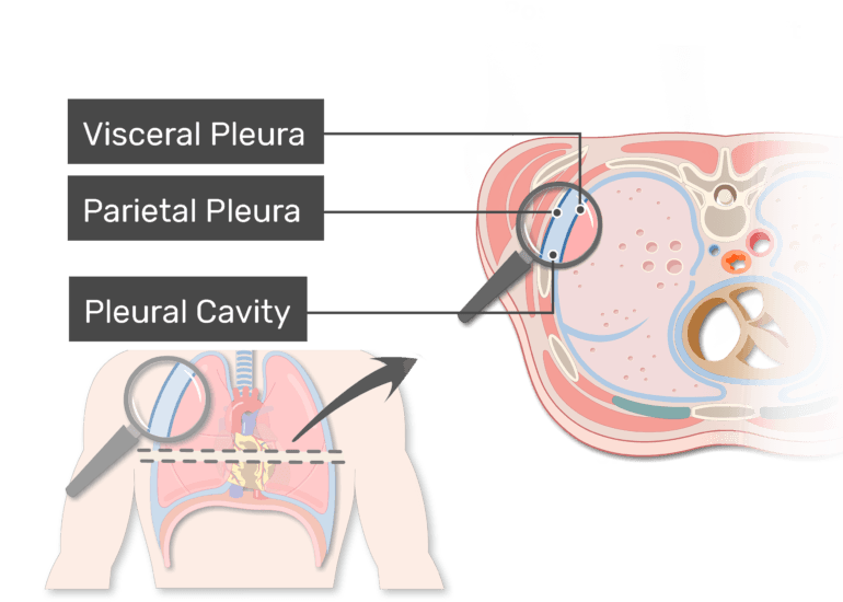
Pleura (or Pleurae) and Pleural Cavity of the Lungs ...
2010 ACCF/AHA/AATS/ACR/ASA/SCA/SCAI/SIR/STS/SVM Guidelines ... Familial aggregation studies of patients referred for repair of thoracic aortic aneurysm and dissection that did not have a genetic defect have indicated that between 11% and 19% of these patients have a first-degree relative with thoracic aortic aneurysms and dissection. 127,130 Patients with a family history of thoracic aortic aneurysm and ...
Sobotta 1906 fig.416 - Transverse section through the upper ...
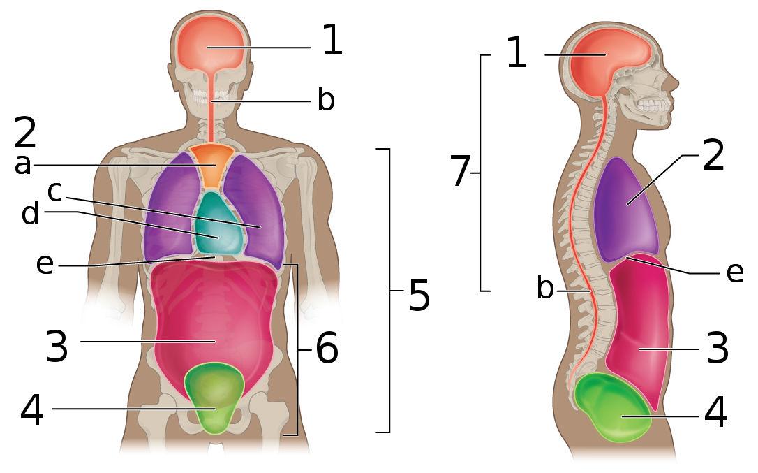
Body Cavities and Membranes: Labeled Diagram, Definitions - EZmed The cranial cavity is the superior portion of the dorsal cavity, as we can see highlighted in red and labeled by the star below. The cranial cavity is enclosed by the cranium or skull, and it houses the brain . The cranial cavity is filled with fluid called cerebrospinal fluid that helps protect and cushion the brain.
File:Body Cavities labeled.png - Wikimedia Commons
Thoracic Cavity - Anatomy | Organs | Functions | 8 Types of Cavities The Thoracic cavity (or chest cavity) is that the chamber of the body of vertebrates that are protected by the pectoral wall ( rib cage and associated skin, fascia, and muscle). The central compartment of the thoracic cavity is the mediastinum.

Thoracic Cavity - Atlas of Anatomy
Label the thoracic cavities.docx - Label the cavities... - Course Hero In the figure above - locate the thoracic cavity. Labelthe structure that separates the thoracic cavity from the abdominopelvic cavity Notice the 4 colors of the thoracic cavity. There are two purple cavities within the thoracic cavity. Labelthem. Identifythe two / three primary structures that lie within the purple cavity. Notice the green cavity.
thoracic
Thoracic Examination - Physiopedia The examiners observe the patient’s thoracic spine region and assess for the presence of deviation from normal including the thoracic spine curvatures in the frontal and sagittal planes. The overall impression of inter-rater reliability for postural observation of kyphosis and label either excessive, normal or decrease range from moderate to ...
1 Thoracic Wall | Radiology Key
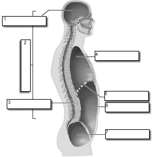
Body Cavities Labeling - The Biology Corner Shows the body cavities from a front view and a lateral view, practice naming the cavity by filling in the boxes. Name: _____ This work is licensed under a Creative Commons Attribution ... Side View: 1. Cranial Cavity 2. Dorsal Cavity 3. Vertebral Canal 4. Thoracic Cavity 5. Diaphragm 6. Abdominal Cavity 7.
TORSOS
Thoracic Lymph Nodes Anatomy, Diagram & Function | Body Maps - Healthline The chest wall thoracic lymph nodes receive drainage from the breasts, arms, pectoral muscles, and other muscles and skin located in the upper section of the chest. Last medically reviewed on July ...
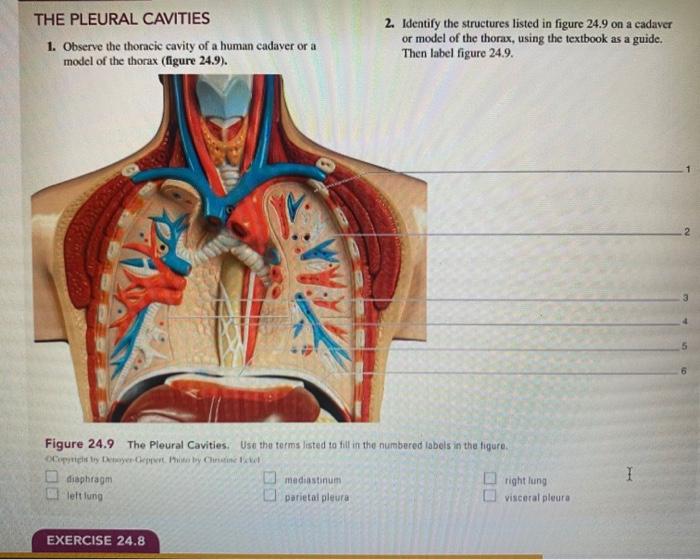
Solved THE PLEURAL CAVITIES 1. Observe the thoracic cavity ...
Body Cavities and Membranes Quiz - Registered Nurse RN The thoracic cavity is located in the ventral cavity. 4. Which landmark separates the thoracic cavity from the abdominopelvic cavity? a. The peritoneum. b. The diaphragm. c. The liver. d. The bladder. The answer is b, the diaphragm. The diaphragm is a muscle that helps us breathe, and it physically separates the thoracic cavity from the ...
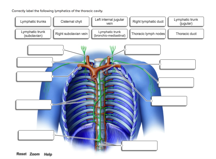
SOLVED: Correctly label the following lymphatics of the ...
A&P 139 Chapter 19 Flashcards | Quizlet Which muscles could assist the diaphragm during inhalation to increase thoracic volume? 2. ... Place each label in the appropriate spot to indicate the muscular activation required to produce the designated volume. ... The mucous membrane lining the nasal cavity warms incoming air. moistens incoming air. entraps dust. all of the above.

Thoracic Cavity Diagram | Quizlet
Human Heart - Diagram and Anatomy of the Heart - Innerbody The heart is located in the thoracic cavity medial to the lungs and posterior to the sternum. On its superior end, the base of the heart is attached to the aorta, Continue Scrolling To Read More Below ... The walls and lining of the pericardial cavity are a special membrane known as the pericardium. Pericardium is a type of serous membrane that ...

File:Body Cavities Frontal view labeled.jpg - Wikimedia Commons
Oxytetracycline (Terramycin®, Liquamycin®) for Dogs and Cats 16-07-2015 · Overview of Oxytetracycline (Terramycin®, Liquamycin®) for Dogs and Cats. Oxytetracycline, also known by the names Terramycin® or Liquamycin®, is an antibiotic that inhibits bacteria by suppressing protein synthesis and growth in dogs and cats.; Oxytetracycline belongs to a general class of drugs known as tetracyclines.

Human Skeleton System Thoracic Skeleton with Label Design ...
Color Diagrams of Insect Organs and Internal Structures Jan 17, 2019 · The first section of the alimentary canal is the foregut or stomodaeum. In the foregut, initial breakdown of large food particles occurs, mostly by saliva. The foregut includes the Buccal cavity, the esophagus, and the crop, which stores food before it passes to the midgut. Once food leaves the crop, it passes to the midgut or mesenteron.

JaypeeDigital | eBook Reader
PDF Body regions, Major body Cavities - Sinoe Medical Association Dorsal Body Cavitywhich houses Cavities the CNS: brain and spinal cord 1). Cranial Cavity 2). Vertebral or spinal cavity •B). Ventral Body Cavity •which houses all other internal body organs 1). Thoracic: •a). Pleural Cavities •b). Pericardial Cavity •c). Mediastinum 2). Abdominopelvic Cavity •a). Abdominal Cavity •b).
Torsos
Body Cavities and Membranes - Anatomy and Physiology Notes The thoracic cavity, also called the chest cavity, sits superior (higher) to the abdominopelvic cavity, and it contains organs such as the heart, lungs, trachea, and esophagus. It can be subdivided into three main portions: The left pleural cavity, which houses the left lung

The Paramedic Shop - The Thoracic Cavity | Facebook
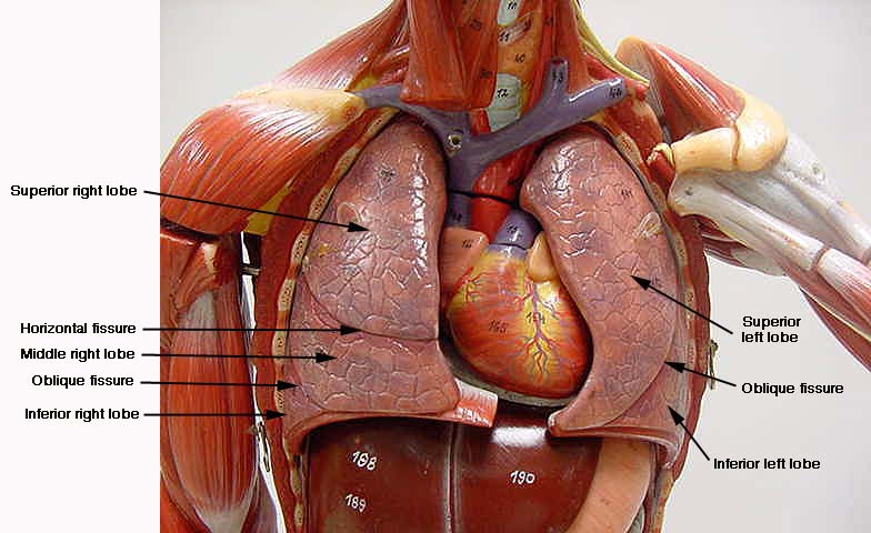
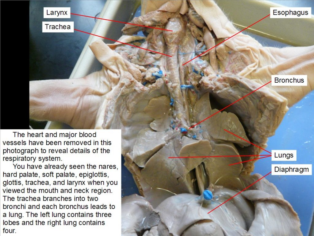
(DOC) Detailed Lesson Plan in Science 10 - Academia.edu The lungs were observed to be pink coloured organ, occupying most of the thoracic cavity, consisting of five portions. The right lung has four lobes cranial, middle, caudal and accessory lobes. The caudal lobe is largest one, while the smallest one is the accessory lobe. The left lung has only one lobe occupied most the left side of thoracic ...
Chapter 11. Fetal Pig Dissection – Anatomy and Physiology 2 ...
Costodiaphragmatic recess - Wikipedia Chest X-ray of a 30-year-old healthy man, with the costodiaphragmatic recess label in red ellipse Front view of thorax , showing the relations of the pleurae and lungs to the chest wall (pleura in blue and lungs in purple)

A&P - Anatomy & Physiology: The Unity of Form and Function ...
Body Cavities Labeling - The Biology Corner Shows the body cavities from a front view and a lateral view, practice naming the cavity by filling in the boxes. Name: _____ This work is licensed under a Creative Commons Attribution ... Side View: 1. Cranial Cavity 2. Dorsal Cavity 3. Vertebral Canal 4. Thoracic Cavity 5. Diaphragm 6.
0514 Anatomy Of Chest Wall And Thoracic Cavity Medical Images ...
Anatomical Body Planes | Science Trends Jan 01, 2019 · The cavities of the body include the dorsal cavity, the cranial cavity, the ventral cavity, the vertebral cavity, the thoracic cavity, and the abdominopelvic cavity. The dorsal cavity is one long continuous cavity that houses portions of the central nervous system including the spinal cord and brain. It is found on the body’s dorsal side.

Membranes and cavities - Human Anatomy - GUWS Medical
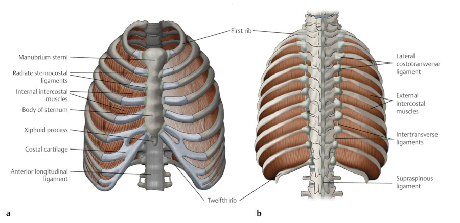
Lab 8: Dissection: Chest Wall, Overview of Thoracic Cavity To download a PDF of this lab guide Goals 1 Clean the thoracic body wall to demonstrate the sternum, ribs, costal cartilages, and intercostal spaces 2 Remove the anterior thoracic wall; Inspect the pleural sacs and mediastinum 3 Open the pleural sacs and define the pleural cavity, parietal pleura, and visceral pleura 4 Remove the right lung

Body Cavities - Body Cavities-Internal Chambers Body cavities ...
thoracic cavity | Description, Anatomy, & Physiology | Britannica thoracic cavity, also called chest cavity, the second largest hollow space of the body. It is enclosed by the ribs, the vertebral column, and the sternum, or breastbone, and is separated from the abdominal cavity (the body's largest hollow space) by a muscular and membranous partition, the diaphragm.
3: The Thorax | Pocket Dentistry
Thoracic Examination - Physiopedia The Thoracic Spine has a complex and often overlooked role within the body. It is a key area of load transfer between the upper and lower body and for rotational movement within the body. Should be assessed and treated as a functional unit including not only the spine but the rib cage.; The thoracic region provides a site for muscle and connective tissue attachments from the …

Anatomy of the Thoracic Wall, Pulmonary Cavities, and ...
Anatomy Chapter 1: Labeling Thoracic Cavity Diagram | Quizlet The cavities surrounding each lung parietal pleura The aspect of the pleura that does not touch the surface of the lung visceral pleura The aspect of the pleura that covers the external surface of the lung The thoracic cavity can be subdivided into... 1. mediastinum 2. left and right pleural cavities 3. pericardial cavity
Anatomy of the Thoracic Wall, Pulmonary Cavities, and ...
Oxytetracycline (Terramycin®, Liquamycin®) for Dogs and Cats Jul 16, 2015 · Oxytetracycline is effective against a wide range of bacteria as well as one-celled (protozoa) organisms. It is effective against bacteria that infect the eyes, oral cavity, bone, respiratory tract, sinuses and blood cells. Oxytetracycline is a prescription drug and can only be obtained from a veterinarian or by prescription from a veterinarian.
Thoracic Cavity Images – Browse 1,613 Stock Photos, Vectors ...
Thoracic Cage Labeling Quiz - PurposeGames.com This is an online quiz called Thoracic Cage Labeling There is a printable worksheet available for download here so you can take the quiz with pen and paper. Your Skills & Rank Total Points 0 Get started! Today's Rank -- 0 Today 's Points One of us! Game Points 13 You need to get 100% to score the 13 points available Actions

Thoracic Cavity - Atlas of Anatomy

Thoracic Images, Illustrations & Vectors (Free) - Bigstock
Merrill's Chapter 10 | Quiz
Thoracic Cavity Photos | Fine Art America

AandP - CH 1 - Body Cavities Labeling - Cranial cavity ...

Thoracic Cage
Pleura (or Pleurae) and Pleural Cavity of the Lungs ...
1.04 Anatomical Terminology- Body Cavities
Post a Comment for "39 label thoracic cavity"