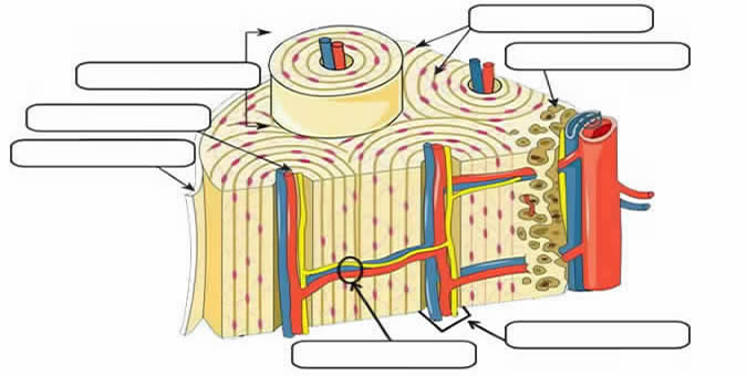
38 label the features associated with the microscopic structure of bone
Microscopic Structure Of Skeleton Muscles - Anatomy Notes The most detail is detected by transmission electron microscopy. Myofibrils, about 1 μm in diameter, are the dominant ultrastructural feature. In longitudinal segments, they appear as ribbons that are interrupted at regular intervals by thin intersecting transverse lines, corresponding to discs in the original cylindrical structure. Solved Trabeculae Bone extracellular matrix Lacuna (space) A - Chegg Question: Trabeculae Bone extracellular matrix Lacuna (space) A FIGURE 12.2 Label the features associated with the microscopic structure of bone. Osteon Lamella Bone extracellular matrix Central canal Lacuna (occupled by osteocyte in living bone) Canaliculi (Victor B. Eichler, Ph.D f und compact bone tissue (200x). APIR
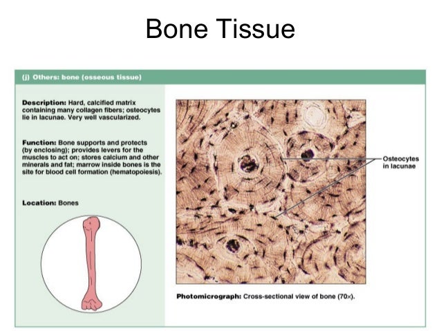
Bone Structure - Anatomy and Physiology - opentextbc.ca The microscopic structural unit of compact bone is called an osteon, or Haversian system. Each osteon is composed of concentric rings of calcified matrix called lamellae (singular = lamella). Running down the center of each osteon is the central canal, or Haversian canal, which contains blood vessels, nerves, and lymphatic vessels.

Label the features associated with the microscopic structure of bone
microscopic bone labeling Flashcards - Quizlet Start studying microscopic bone labeling. Learn vocabulary, terms, and more with flashcards, games, and other study tools. Types of Bone Cells | Osteoclasts, Osteoblasts, & Osteocytes Both the compact and spongy bone tissues are composed of 3 main types of bone cells. These bone cells have distinct features, structure, and considered essential functions. These bone cells are Osteoclasts, Osteoblasts, and Osteocytes. These bone cells are embedded in the matrix of bony tissue and perform many vital functions. Solved us Trabeculae CSLS ea 8 Bone extracellular matrix 10- | Chegg.com A A Question: us Trabeculae CSLS ea 8 Bone extracellular matrix 10- Lacuna (space) FIGURE 12.2 Label the features associated withthe microscopic structure of bone. A A This problem has been solved! See the answer Show transcribed image text Expert Answer 100% (2 ratings) 6. Perforating canal 5. Arte … View the full answer
Label the features associated with the microscopic structure of bone. Structure of Bone Tissue | SEER Training Spongy (cancellous) bone is lighter and less dense than compact bone. Spongy bone consists of plates ( trabeculae) and bars of bone adjacent to small, irregular cavities that contain red bone marrow. The canaliculi connect to the adjacent cavities, instead of a central haversian canal, to receive their blood supply. PDF Bones and Bone Structure - Palm Beach State College Section 1: Introduction to the Structure and Growth of Bones Learning Outcomes 6.1 Describe the two main divisions of the skeleton, and list the major functions of the skeletal system. 6.2 Classify bones according to their shapes, identify the major types of bone markings, and explain the functional significance of bone markings Bone Structure - Anatomy & Physiology - University of Hawaiʻi The microscopic structural unit of compact bone is called an osteon, or Haversian system. Each osteon is composed of concentric rings of calcified matrix called lamellae (singular = lamella). Running down the center of each osteon is the central canal, or Haversian canal, which contains blood vessels, nerves, and lymphatic vessels. 6.3 Bone Structure - Anatomy & Physiology If you look at compact bone under the microscope, you will observe a highly organized arrangement of concentric circles that look like tree trunks. Each group of concentric circles (each "tree") makes up the microscopic structural unit of compact bone called an osteon (this is also called a Haversian system).
Bone marrow: Histology, types and features | Kenhub The bony skeleton that supports the human body and facilitates locomotion has an intricate microarchitecture of its own. The cavities created by the trabecular arrangement of the core of the bones are occupied by a mixture of blood cells across a large spectrum of development, and adipocytes. 6.3 Bone Structure - Anatomy and Physiology | OpenStax Bone Markings. The surface features of bones vary considerably, depending on the function and location in the body. Table 6.2 describes the bone markings, which are illustrated in (Figure 6.10). There are three general classes of bone markings: (1) articulations, (2) projections, and (3) holes. figure 8.2 A (microscopic features of bone) Flashcards | Quizlet Start studying figure 8.2 A (microscopic features of bone). Learn vocabulary, terms, and more with flashcards, games, and other study tools. Bones: Types, structure, and function - Medical News Today Bones provide a frame to support the body. Muscles, tendons, and ligaments attach to bones. Without anchoring to bones, muscles could not move the body. Some bones protect the body's internal...
Structure and Function of the Haversian System Explained ... - Bodytomy The terms 'Haversian system' or 'osteon' refer to the basic cylindrical-shaped structural unit of a compact bone, which in turn forms a substantial part of the structure of the long bones of the human body. The osteons are closely packed, with osteocytes lined up in concentric rings. This imparts a hard and dense texture to the compact ... Lab Report 12 Bone Structure and Classification - Quizlet Melanin is used to protect the cell's nucleus from UV radiation. Figure 12.2 features associated with the microscopic structure of bone. 1. Spongy Bone 2. Compact Bone 3. Osteon 4. Periosteum 5. Central Canal 6. Perforating Canal 7. Blood Vessels 8. Nerve 9. Canaliculus 10. Osteocyte A bone that is platelike is classified as a (n) ________ bone. PDF Bone Structure Description Lab - burkesci.weebly.com Label the femur (an example of a long bone) below with the following: Memorize the structures by quizzing each other in pairs Label the following features associated with the microscopic structure of bone: (use twice) 1) Blood vessels (use twice) 2) Canaliculi 3) Compact bone (use twice) 4) Endosteum 5) Haversian canal Skeletal System - Labeled Diagrams of the Human Skeleton Each bone is a complex living organ that is made up of many cells, protein fibers, and minerals. The skeleton acts as a scaffold by providing support and protection for the soft tissues that make up the rest of the body. The skeletal system also provides attachment points for muscles to allow movements at the joints.

Epithelium, cells,tissues & histology
Exercise 8.2 - Microscopic Features of Bone - Photos - Quizlet Taken from Exercise 8 : Bone Structure and Function Pages: 98 Microscopic features of bone Exercise 8.2 - Microscopic Features of Bone - Photos study guide by lumina8 includes 14 questions covering vocabulary, terms and more. Quizlet flashcards, activities and games help you improve your grades.
Fruit: Microscopic Structure Of Spongy Bone
10.2 Skeletal Muscle - Anatomy & Physiology The sarcomere is the smallest functional unit of a skeletal muscle fiber and is a highly organized arrangement of contractile, regulatory, and structural proteins. It is the shortening of these individual sarcomeres that lead to the contraction of individual skeletal muscle fibers (and ultimately the whole muscle).
Epithelium, cells,tissues & histology
Structure Of Bone - Human Anatomy - GUWS Medical Reexamine the microscopic structure of bone tissue by observing a prepared microscope slide of ground compact bone. Use the figures of bone tissue in a textbook to locate the following features: osteon (Haversian system) osteonic canal (Haversian canal) lamella lacuna (small chamber for an osteocyte) canaliculus Critical Thinking Application

Solved: Trabeculae Bone Extracellular Matrix Lacuna (space... | Chegg.com
Compact Bone Structure - Biology Dictionary Compact bone, also called cortical bone, is the hard, stiff, smooth, thin, white bone tissue that surrounds all bones in the human body. It is also called osseous tissue or cortical bone and it provides structure and support for an organism as part of its skeleton, in addition to being a location for the storage of minerals like calcium.About 80% of the weight of the human skeleton comes from ...

Pin on Brain Stuff!
PDF scripTower - link Figure 12.2 Figure 12.3 Trabeculae Bone extracellular Lacuna (space) Label the features associated with the microscopic structure of bone. Osteon Lamella extracellular matrix Central canal Lacuna (occupied by osteocyte in living bone) Canaliculi Micrograph Of ground compact bone tissue (200>0. 87
Notes Ch 7 (Skeleton)
HOLES+LAB+12.pdf - -"~ ~- -~-~=~=.= ') . Laboratory 2 Exercise Bone ... use figure 12.3 of bone tissue to locate the following features: osteon (haversian system)-cylinder-shaped unit central canal (haversian canal) -contains blood vessels and nerves lacuna-small chamber for an osteocyte bone extracellular matrix-collagen and calcium phosphate lamella-concentric ring of matrix around central canal canaliculus-minute …
Post a Comment for "38 label the features associated with the microscopic structure of bone"