44 label the photomicrogram of the trachea.
Free Automated Malware Analysis Service - powered by Falcon Sandbox ... Submit malware for free analysis with Falcon Sandbox and Hybrid Analysis technology. Hybrid Analysis develops and licenses analysis tools to fight malware. ChartNet Tech o, o= +, o, o> (- o oI X 2Ð o? Ži 1 o‚ Þ , o@ Ü * ,Z† sS * sU * sM * sP * sc * se * s] * s_ * sg * s[ *.( € * {A * {B * {C * (' * (' *ž {D {E {D {F oG {H ...
Solved Respiratory Lab Worksheet Saved Help Save & Exit - Chegg See the answer Label the photomicrograph of the trachea Show transcribed image text Expert Answer 100% (4 ratings) Transcribed image text: Respiratory Lab Worksheet Saved Help Save & Exit Sbmit Label the photomicrogram of the trachea. Connect 0.23 points Cartilage Print Epithelium References Submucosa Perichondrium Lamina propria Reset Zoom
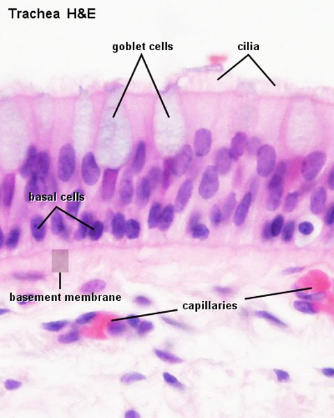
Label the photomicrogram of the trachea.
(Get Answer) - Determine the angle of i, r and q this is reflection of ... Determine the angle of i, r and q this is reflection of light General Structure of Mucosa Label the structures that comprise the respiratory tract mucosa (mucous membrane). ... lung Segmental bronchus Trachea prey="" 11="" of="" 46="" next=""> Trachea histology of respiratory system low power Label the photomicrogram of the trachea. A&P 2 Lab Unit 2 Flashcards | Quizlet Label the photomicrogram of the trachea. Identify these structures in the left-sided midsagittal view of the superior portion of the lower respiratory system. Identify the anatomical structures shown in the picture of the thorax. Identify the anatomical structures shown in a lateral view of the left lung. Solved Label the photomicrogram of the trachea. Cilia Lamina | Chegg.com Question: Label the photomicrogram of the trachea. Cilia Lamina propria Submucosa Cilia Basement membrane Submucosa Epithelium Basement membrane Lamina propria Epithellum This problem has been solved! See the answer why is it telling me that those are wrong? Show transcribed image text Expert Answer 100% (6 ratings)
Label the photomicrogram of the trachea.. Histology of trachea and lung - SlideShare 1. HISTOLOGY OF TRACHEA AND LUNG Dr.ushakannan,Asst.professor. 2. RESPIRATORY SYSTEM Conducting Part- responsible for passage of air and conditioning of the inspired air. Examples:nasal cavities,pharynx, trachea, bronchi and their intrapulmonary continuations. Respiratory Part-involved with the exchange of oxygen and carbondioxide between blood ... The Trachea (Human Anatomy): Picture, Function, Conditions, and More The trachea, commonly known as the windpipe, is a tube about 4 inches long and less than an inch in diameter in most people. The trachea begins just under the larynx (voice box) and runs down... BIO208 Lab Practical 2 - 10/6/2019 Lab Practical 2 Home - Course Hero 10/6/2019 Lab Practical 2 Question Label the structure with a "star" symbol beside it. 4 Incorrect Mark 0.00 out of 1.00 Answer: trachea The correct answer is: larynx (based on the document attached to the question as reference) Histology, Alveolar Cells - StatPearls - NCBI Bookshelf Alveoli represent the most distal portion of the respiratory tract. There are approximately 500 million alveoli in the human body.[1] Each alveolus is separated from the other by an alveolar septum, which contains the pulmonary capillaries participating in gas exchange and connective tissue.
Trinidad State College Home Page Trinidad State is a Hispanic-Serving Institution (HSI) HSI is defined in federal law (the Higher Education Opportunity Act, Title V, 2008) as an accredited, degree-granting, public or private nonprofit institution of higher education with 25% or more total undergraduate Hispanic full-time equivalent (FTE) student enrollment. Anatomy A215 Virtual Microscopy - Indiana University Bloomington Anatomy A215 Virtual Microscopy Each alveolus is a small air space surrounded by an extensive capillary network. The epithelium which lines the alveoli is an extremely thin simple squamous in close proximity to the capillary walls. Alveoli make up the major part of the lung and give it its sponge-like appearance under the microscope. Label The Photomicrograph Of The Lung : 4 Chloro Dl Phenylalanine ... Label The Photomicrograph Of The Lung : 4 Chloro Dl Phenylalanine Protects Against Monocrotaline Induced Pulmonary Vascular Remodeling And Lung Inflammation. The lower respiratory system., put the following layers of the trachea in order from superficial to deep., label the structures of the upper respiratory . A&P 139 Chapter 19 Flashcards | Quizlet Label the photomicrogram of the lung. Label the photomicrogram of the trachea. Cricoid. Which of these laryngeal cartilages is single? Label these structures of the upper respiratory system. tidal volume. The volume of air that enters (or leaves) during a single respiratory cycle is the.
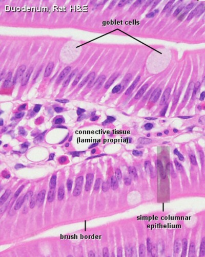
Labeled diagram of the lungs/respiratory system. - SERC View Original Image at Full Size. Labeled diagram of the lungs/respiratory system. Image 37789 is a 1125 by 1408 pixel PNG Uploaded: Jan10 14. Last Modified: 2014-01-10 12:15:34 (Lee Ann C. Golper) Medical Speech-Language Pathol (BookFi) [Lee_Ann_C._Golper]_Medical_Speech-Language_Pathol(BookFi).pdf - Free ebook download as PDF File (.pdf), Text File (.txt) or read book online for free. Medical Speech-Language Pathology: A Desk Reference, Third Edition ... • Avoid personal comments or assigning blame for a problem. • Avoid excessive spacing within and between notes. • If notes exceed one page, write "continued on pg 2" at the bottom of the first page and then "Speech Path Note continued from pg 1" at the top of the second page. • Use descriptions in place of labels. Lab 2: Microscopy and the Study of Tissues - UW-La Crosse The lining of the trachea consists of a type of tissue called pseudostratified (ciliated) columnar epithelium. This single layer of ciliated cells appears stratified because the cells vary in their thickness and because their nuclei are located at different levels. 2 - Pseudostratified columnar epithelium (close-up view) Ciliated border
File:Trachea histology 01.jpg - Embryology
Can you label the lungs? Quiz - PurposeGames.com This is an online quiz called Can you label the lungs? There is a printable worksheet available for download here so you can take the quiz with pen and paper. From the quiz author. Labeling the lungs. This quiz has tags. Click on the tags below to find other quizzes on the same subject. lungs. respiratory system.

Gallery For Trachea Histology | Trachea, Nursing labs, Basement membrane
Solved Label the photomicrogram of the trachea. Cilia Lamina | Chegg.com Question: Label the photomicrogram of the trachea. Cilia Lamina propria Submucosa Cilia Basement membrane Submucosa Epithelium Basement membrane Lamina propria Epithellum This problem has been solved! See the answer why is it telling me that those are wrong? Show transcribed image text Expert Answer 100% (6 ratings)

Anatomy And Physiology Archive | October 25, 2017 | Chegg.com
A&P 2 Lab Unit 2 Flashcards | Quizlet Label the photomicrogram of the trachea. Identify these structures in the left-sided midsagittal view of the superior portion of the lower respiratory system. Identify the anatomical structures shown in the picture of the thorax. Identify the anatomical structures shown in a lateral view of the left lung.
Foundations - Histology Epithelia and Skin - Embryology
(Get Answer) - Determine the angle of i, r and q this is reflection of ... Determine the angle of i, r and q this is reflection of light General Structure of Mucosa Label the structures that comprise the respiratory tract mucosa (mucous membrane). ... lung Segmental bronchus Trachea prey="" 11="" of="" 46="" next=""> Trachea histology of respiratory system low power Label the photomicrogram of the trachea.

lung histology labeled - bronchiole, alveolar duct, alveoli | lab ...

Anatomy of the Respiratory System | Respiratory, Histology slides ...
Post a Comment for "44 label the photomicrogram of the trachea."